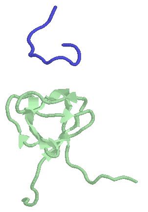
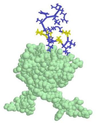
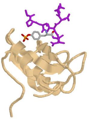
Grb2, or growth factor receptor-bound protein 2, is an adaptor protein involved in signal transduction [1]. It contains one SH2 protein domain and two SH3 protein domains. The SH2 domain (Src homology 2, figure 1) binds to phorsphorylated tyrosine residues in a protein interaction partner, while the SH3 (Src homology 3) domain binds to the proline rich surfaces with a PXXP binding motif in a corresponding protein interaction partner or peptide (Figure 1) [2, 3, 4]. One example of a known pathway Grb2 is involved in is the induced gene expression by insulin. In the cell, Grb2 relays the message from IRS-1, when its tyrosine residues are phosphorylated, to Sos, which contains a proline rich region.
|
a)
|
b)
|
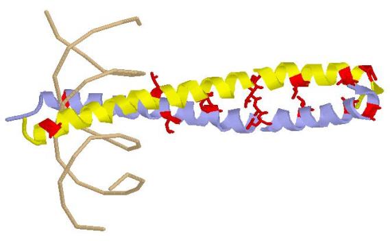
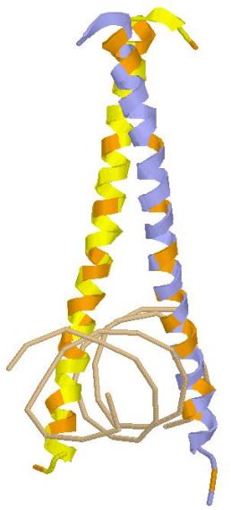
GCN4 is a transcriptional activator protein [5]. Although it was first characterized to positively regulate expressed genes during amino acid starvation, microarray experiments have found that GCN4 may be involved in additional processes such as purine biosynthesis, organelle biosynthesis, and multiple stress responses. GCN4 is a homodimer that forms a leucine zipper, two alpha helices coiled around each other with each helical hydrophobic amino acid residues assembled near the surface where the two helices interact [4]. The leucine zipper is a common DNA-binding motif where leucine residues of each helix recur every seventh position on the hydrophobic side of the structure (Figure 3). The structural domain name, ōleucine zipper,ö is actually a misnomer as the leucine residues are only lined up against one another and do not really ōinterdigitateö as the name implies. Usually, regulatory proteins with leucine zippers also contain a DNA-binding domain, characteristically containing ample amounts of basic amino acid residues (Lys or Arg) that can interact with the negatively charged DNA backbone (Figure 4).
[1] NCBI. ōGene: GRB2 growth factor receptor-bound protein 2 [Homo Sapiens].ö
National Institutes of Health. National Library of Medicine. April 16, 2007.
http://www.ncbi.nlm.nih.gov/entrez/query.fcgi?db=gene&cmd=Retrieve&dopt=Graphics&list_uids=2885
[2] Pfam. ōSH2 Domain.ö April 16, 2007. http://www.sanger.ac.uk/cgi-bin/Pfam/getacc?PF00017
[3] Pfam. ōSH3 Domain.ö April 16, 2007. http://www.sanger.ac.uk/cgi-bin/Pfam/getacc?PF00018
[4] Nelson, D. and Cox, M. Lehnineger Principles of Biochemistry. New York:
W. H. Freeman and Company. 2005. pp. 429, 1090-1091.
[5] Saccharomyces Genome Database. ōGCN4 Basic Information.ö Stanford
University. April 16, 2007. http://db.yeastgenome.org/cgi-bin/locus.pl?locus=GCN4